


What Is an Eye OCT Scan and What Is It Used For?
An eye OCT scan, also known as optical coherence tomography, is a diagnostic test that uses light waves to produce detailed images of the different layers of the retina and optic nerve.
It can be compared to a scan of the inside of the eye. The test allows us to view structures that cannot always be examined in the same level of detail during a conventional eye examination.
An eye OCT scan is mainly used to:
- Examine the retina and macula.
- Assess the optic nerve.
- Detect and monitor glaucoma.
- Identify macular changes.
- Assess macular oedema.
- Monitor retinal diseases.
- Compare changes in the eye over time.
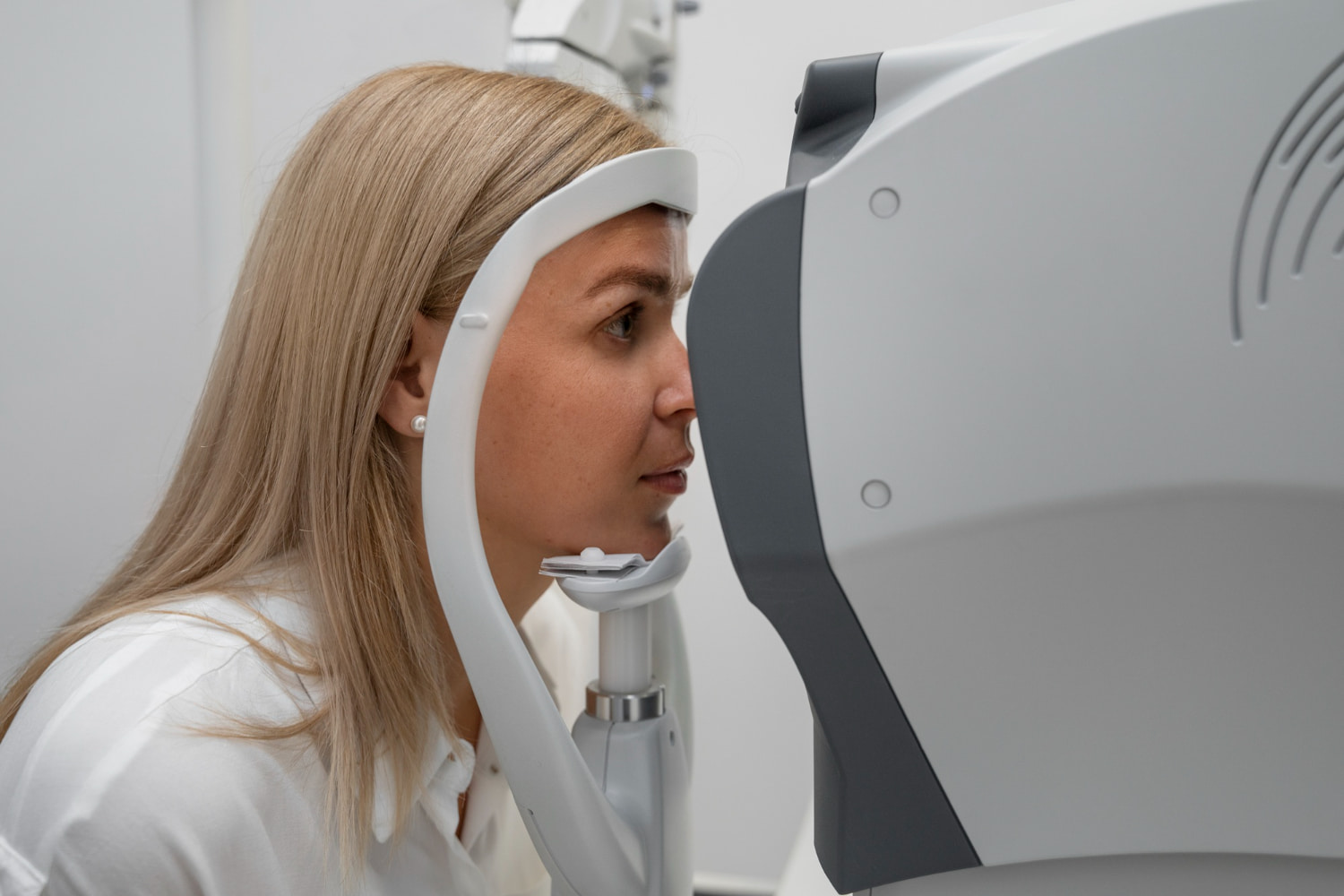
It is a quick, non-invasive and usually painless test. You simply need to keep your gaze fixed for a few seconds while the equipment captures the images.
OCT does not replace a comprehensive eye examination. Its findings must be interpreted alongside visual acuity, intraocular pressure, examination of the back of the eye and any other tests that may be indicated.
What Does OCT Stand For?
OCT stands for optical coherence tomography.
It is an imaging technology that uses light to obtain cross-sectional views of the eye’s structures.
The scan allows the ophthalmologist to examine:
- The different layers of the retina.
- The thickness of the macula.
- The retinal nerve fibre layer.
- The optic nerve head.
- Certain structures at the front of the eye.
The images produced allow measurements to be taken and the findings to be compared with previous scans.
This is particularly useful for chronic conditions, where it is important to know whether a change remains stable or progresses over time.
How Does an Eye OCT Scan Work?
OCT directs a beam of light towards the eye’s structures and analyses how that light is reflected by the different tissues.
Each layer has different characteristics. The equipment processes this information and creates cross-sectional images, similar to microscopic slices, without touching or operating on the eye.
The scan can:
- Show the internal structure of the retina.
- Measure the thickness of different layers.
- Locate fluid build-up.
- Detect changes in the optic nerve.
- Compare the two eyes.
- Assess changes between appointments.
Although the equipment provides maps, measurements and statistical comparisons, a diagnosis should not be based solely on the colours or values in the automated report. The images must be reviewed and interpreted by the professional in the context of each patient’s clinical findings.
Which Structures Can an OCT Scan Examine?
Macular OCT
The macula is the central area of the retina responsible for fine, detailed vision.
It allows us to:
- Read.
- Recognise faces.
- See fine details.
- Drive.
- Carry out precision work.
Macular OCT makes it possible to assess the shape and thickness of its different layers.
It can show:
- Fluid build-up.
- Inflammation.
- Traction on the retina.
- Membranes.
- Macular holes.
- Degenerative changes.
Optic Nerve OCT
The optic nerve carries visual information from the eye to the brain.
OCT can assess the optic nerve head and measure the thickness of the surrounding retinal nerve fibre layer.
This assessment is particularly important for:
- Glaucoma.
- Suspected glaucoma.
- Ocular hypertension.
- Optic nerve abnormalities.
- Monitoring certain forms of neurological damage.
Anterior Segment OCT
Some OCT systems can also examine the front of the eye.
They can assess:
- The cornea.
- The anterior chamber angle.
- The iris.
- The depth of certain structures.
Whether it is indicated depends on the condition being investigated and the type of equipment available.
What Is an Eye OCT Scan Used For?
OCT can be used to detect, document and monitor a range of conditions.
Its main aims are to:
- Obtain detailed images of the retina and optic nerve.
- Detect changes that may not be apparent during an initial examination.
- Measure the thickness of certain structures objectively.
- Compare the findings with previous scans.
- Assess whether a condition is progressing.
- Check the response to treatment.
- Help plan the patient’s follow-up.
Its specific usefulness always depends on the reason for the appointment and the findings from the examination.
Which Conditions Can Be Assessed with OCT?
1. Glaucoma
Glaucoma can cause progressive loss of nerve fibres and damage the optic nerve.
OCT can measure:
- The retinal nerve fibre layer.
- The ganglion cell complex.
- Certain characteristics of the optic nerve.
These measurements help to:
- Detect suspicious signs.
- Establish an initial baseline.
- Compare findings over time.
- Assess possible signs of progression.
However, glaucoma cannot be diagnosed with an OCT scan alone.
It may also be necessary to assess:
- Intraocular pressure.
- The visual field.
- The clinical appearance of the optic nerve.
- Corneal thickness.
- The drainage angle.
- Personal and family history.
2. Age-related macular degeneration
Macular degeneration affects the central area of the retina.
OCT can show:
- Changes in the retinal layers.
- Deposits beneath the retina.
- Fluid build-up.
- Changes in the retinal pigment epithelium.
- Signs of atrophy.
It can also be used to monitor the response to certain treatments.
3. Macular oedema
Macular oedema is a build-up of fluid within or beneath the central retina.
It may be associated with:
- Diabetes.
- Retinal vascular occlusions.
- Inflammation.
- Eye surgery.
- Other retinal diseases.
OCT can locate the fluid and measure the thickness of the macula.
4. Diabetic retinopathy
Diabetes can damage the blood vessels in the retina.
OCT is particularly useful when diabetic macular oedema is suspected or when changes in the central retina need to be monitored.
The scan does not replace examination of the back of the eye or any other tests that may be indicated.
5. Epiretinal membrane
An epiretinal membrane is a thin layer of tissue that can form on the surface of the macula.
It can cause:
- Blurred vision.
- Distorted lines.
- Difficulty reading.
- Differences in vision between the two eyes.
OCT can show the membrane and whether it is distorting the retina.
6. Macular hole
A macular hole is an opening that develops in the central retina.
OCT helps to:
- Confirm its presence.
- Determine its size.
- Assess its configuration.
- Plan treatment.
- Monitor subsequent changes.
7. Vitreomacular traction
In some patients, the vitreous remains attached to the macula and pulls on it.
OCT shows this relationship in great detail and can reveal whether it is affecting the anatomy of the retina.
8. Retinal detachments and other retinal changes
OCT can provide information about certain retinal changes and help locate structural abnormalities.
However, if you experience flashes, a sudden increase in floaters or a curtain across your field of vision, it is essential to have an urgent dilated eye examination.
9. Optic nerve disorders
In addition to glaucoma, OCT can be used as a supplementary test for certain optic nerve disorders.
The findings must be interpreted alongside:
- The medical history.
- Visual acuity.
- Colour vision.
- The pupil examination.
- The visual field test.
- Other neurological tests when indicated.
How Is an Eye OCT Scan Performed?
The test usually involves the following steps:
- You sit in front of the equipment.
- You rest your chin and forehead on the supports.
- You look at a target light indicated by the professional.
- You should try to keep your eye open and your gaze fixed.
- The equipment performs several light scans.
- The professional checks the quality of the images.
- Any image that is not sufficiently clear may be repeated.
No instrument is placed inside the eye.
The test usually takes only a few minutes, although it may take a little longer if images need to be repeated or different areas examined.
Does an Eye OCT Scan Hurt?
No. OCT is non-invasive and normally painless.
You may notice:
- The light from the equipment.
- The need to keep your gaze fixed.
- Mild tiredness if the scan has to be repeated several times.
It does not use ionising radiation or involve direct contact with the inside of the eye.
Do the Pupils Need to Be Dilated?
Not always.
Suitable images can be obtained without dilation in many patients.
Dilation may be needed when:
- The pupil is small.
- The image quality is insufficient.
- Cataracts or other opacities are present.
- A comprehensive examination of the back of the eye is also needed.
After dilation, near vision may be blurred and sensitivity to light may increase for a few hours.
How Long Does an Eye OCT Scan Take?
Each image takes only a few seconds to capture.
The total time depends on:
- The number of areas that need to be examined.
- How well you can keep your gaze fixed.
- Whether any scans need to be repeated.
- The clarity of the ocular media.
It is usually completed within a few minutes.
Do You Need to Prepare for an OCT Scan?
It does not generally require any special preparation.
We recommend that you:
- Bring any previous reports or test results if they were carried out at another clinic.
- Tell us about any previous eye conditions.
- Tell us about any treatments you are currently using.
- Bring sunglasses in case your pupils need to be dilated.
You do not usually need to fast or stop your regular medication unless specifically advised.
What Do the Colours on an OCT Scan Mean?
OCT reports often use colours to represent thickness measurements or compare them with a reference database.
Depending on the equipment, the report may show:
- Green areas.
- Yellow areas.
- Red areas.
- Thickness maps.
- Progression charts.
A red result does not confirm a condition by itself, just as a green result does not guarantee that the eye is healthy.
The colours can be affected by:
- Individual anatomy.
- High short-sightedness.
- Segmentation errors.
- Poor image quality.
- Patient movement.
- The presence of other eye conditions.
That is why the scan must be interpreted as a whole rather than relying on the colour coding alone.
Does an Abnormal OCT Scan Mean I Have Glaucoma?
Not necessarily.
An OCT scan can show reduced thickness of the nerve fibre layer for several reasons.
Abnormal results can also be caused by:
- High short-sightedness.
- Anatomical features of the optic nerve.
- Retinal conditions.
- Poor image quality.
- Errors in the automated analysis.
A glaucoma diagnosis requires all the findings to be assessed together.
Does a Normal OCT Scan Rule Out Every Eye Condition?
No.
An OCT scan can appear normal even when there is:
- A refractive error.
- A cataract.
- Dry eye.
- A visual field defect that is not yet detectable structurally.
- A condition at a stage that is not visible on this test.
- A problem outside the scanned area.
OCT is a complementary test and must be interpreted as part of a comprehensive eye examination.
How Often Should an OCT Scan Be Repeated?
There is no single schedule that is right for every patient.
It may be carried out:
- Once, to investigate a specific symptom.
- Annually, as part of ongoing monitoring.
- Every few months if there is a risk of progression.
- Before and after certain treatments.
- More frequently when the specialist needs to assess rapid changes.
How often it is needed depends on:
- The condition being assessed.
- Its severity.
- The rate of progression.
- The treatment being used.
- The results of other tests.
How Does an OCT Scan Differ from a Fundus Examination?
A fundus examination allows the retina, macula, blood vessels and optic nerve to be viewed directly.
An OCT scan provides cross-sectional images of the internal layers and quantitative measurements.
The two tests provide different and complementary information.
| Test | Main information |
| Fundus examination | Overall appearance of the retina, macula, blood vessels and optic nerve |
| OCT | Cross-sectional images and measurements of the retinal layers and optic nerve |
| Visual field test | Function of central and peripheral vision |
| Fundus photography | Colour photograph of the back of the eye |
How Does an OCT Scan Differ from a Visual Field Test?
An OCT scan primarily assesses structure.
A visual field test assesses function: which areas the patient can see while keeping their gaze fixed.
Both are commonly used to monitor glaucoma because:
- OCT measures the nerve tissue.
- A visual field test shows how the damage affects vision.
You can find out more about our ophthalmic diagnostic tests.
Limitations of Eye OCT
Although it is a very useful test, OCT does have some limitations.
Image quality may be reduced by:
- Advanced cataracts.
- Corneal opacities.
- Eye movements.
- Difficulty keeping the gaze fixed.
- Very small pupils.
- Blinking during image acquisition.
- Very high short-sightedness.
Automated systems may also identify some of the retinal layer boundaries incorrectly.
The ophthalmologist must check the scan quality and make sure the measurements truly reflect the patient’s anatomy.
Frequently Asked Questions About Eye OCT
What Is an Eye OCT Scan?
It is a non-invasive imaging test that uses light to produce detailed cross-sectional images of the retina and optic nerve.
What Is an OCT Scan Used For?
It is used to detect, measure and monitor changes affecting the macula, retina and optic nerve.
Does an Eye OCT Scan Hurt?
No. It is normally a painless test with no direct contact with the eye.
How Long Does It Take?
The images can usually be captured in just a few minutes.
Does OCT Use Radiation?
It does not use X-rays or ionising radiation. It works with light waves.
Do the Pupils Need to Be Dilated?
Not always. Dilation may be needed if the pupil is small or the image quality is not good enough.
Can OCT Detect Glaucoma?
It can reveal changes in the retinal nerve fibre layer and support diagnosis and monitoring, but it should not be used in isolation.
Can OCT Detect Retinal Problems?
Yes. It can show oedema, membranes, holes, traction and other structural changes.
Does a Normal OCT Scan Mean My Eyes Are Healthy?
Not necessarily. Some eye conditions and vision problems require other tests.
Can I Drive After an OCT Scan?
Yes, unless your pupils have been dilated and your vision is temporarily blurred or you are particularly sensitive to light.
How Often Should It Be Repeated?
This depends on the condition and the risk of progression. Your ophthalmologist will recommend the appropriate interval.
Who Interprets an OCT Scan?
The ophthalmologist interprets the images alongside the rest of the eye examination and the patient’s medical history.
A Detailed Scan Must Be Interpreted as Part of a Complete Eye Examination
Eye OCT makes it possible to examine and measure the retinal layers and certain structures of the optic nerve in great detail.
It is particularly useful for diagnosing and monitoring glaucoma, macular conditions, retinal oedema and other eye disorders.
At Centro Oftalmológico del Llano, we use OCT as part of an assessment tailored to each patient, considering the results alongside visual acuity, eye pressure, the fundus examination and any other tests required.
You can find out more about our ophthalmic diagnostic tests.